In 2026, animal surgery safety depends on disciplined preparation, not technology alone. The question is practical: how to ensure safety during animal surgery when every patient responds differently? A small dog may become hypothermic under bright operating lights. A cat may hide pain until recovery is already unstable. These details demand structured decisions before, during, and after anesthesia.
The AAHA Anesthesia and Monitoring Guidelines emphasize individualized assessment, continuous monitoring, trained personnel, and documented recovery care. RECOVER’s evidence-based veterinary CPR guidance also supports clear team roles, rapid recognition, and rehearsed emergency responses. These professional standards provide a reliable foundation, but they are not a substitute for clinical judgment. Equipment alarms can be missed. Checklists can become routine paperwork. That is uncomfortable, but important.
Safety starts with a complete history, physical examination, laboratory review, and risk classification. Teams should confirm patient identity, procedure, allergies, fasting status, consent, and blood availability when appropriate. During surgery, temperature, oxygenation, ventilation, circulation, anesthetic depth, and pain should be assessed continuously. An assistant should record trends rather than isolated readings. After closure, monitoring must continue until the animal maintains stable breathing, circulation, temperature, and responsiveness.
Small errors matter.
The 2026 operating room should combine validated monitors, simulation training, antimicrobial stewardship, and honest incident review. A near miss is not a personal failure. It is evidence that a process needs improvement. Reliable veterinary teams learn from those moments, communicate clearly, and keep the animal’s welfare above speed or convenience.
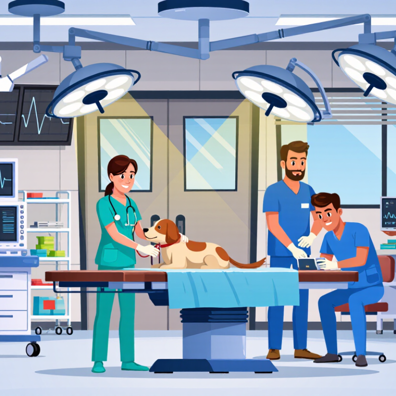
Preoperative risk assessment begins with the animal, not the procedure. Record its species, breed, age, weight, temperament, and previous anesthetic response. Examine hydration, breathing, heart sounds, mucous membranes, pain, and body temperature. A quiet cat may hide serious illness. Small details matter.
Review current medicines, allergies, vaccination history, and recent changes in appetite or behavior. Blood tests can reveal anemia, infection, kidney stress, or electrolyte imbalance. Imaging may clarify fractures, foreign material, or abdominal disease. Select tests according to the patient’s condition, not habit. A healthy young animal may need less testing, while an older patient may require broader evaluation. I have learned that rushed histories create avoidable surprises. No checklist catches everything.
Tips: Confirm fasting instructions and document the last meal. Prepare a weight-based anesthetic plan, intravenous access, monitoring equipment, warming support, and recovery space. Assign one person to monitor vital signs continuously. Discuss pain control before surgery, including rescue options if pain exceeds expectations. Plan for bleeding, difficult airways, unexpected findings, or delayed recovery. Confirm that necessary staff, instruments, oxygen, and emergency medicines are ready. Recheck patient identity and the intended surgical site before sedation. Ask the owner clear questions, and explain realistic risks without promising certainty. A second review can expose a missed detail.
| Risk-Assessment Dimension | Key Data to Collect | Planning Considerations | Suggested Safety Trigger | Required Documentation |
|---|---|---|---|---|
| Physical status and urgency | Species, age, body condition, hydration, temperature, heart rate, respiratory rate, mucous membranes, pain score, and emergency status. | Assign a physical-status category such as ASA 1–5 and identify whether the procedure is elective, urgent, or emergent. | Postpone elective surgery when unstable perfusion, uncontrolled pain, severe dehydration, or respiratory compromise is present. | Pre-anesthetic examination, risk category, urgency level, and stabilization plan. |
| Medical history and medication review | Previous anesthetic reactions, current medicines, supplements, allergies, recent illness, vaccination status, and prior surgeries. | Assess effects on bleeding, blood pressure, renal function, glucose control, sedation, and drug interactions. | Clarify any medication that may increase anesthetic or bleeding risk before consent and drug selection. | Updated medication list, allergy record, previous anesthetic-event notes, and clinician instructions. |
| Airway and respiratory assessment | Airway anatomy, brachycephalic features, coughing, exercise intolerance, respiratory effort, thoracic findings, and aspiration risk. | Prepare appropriately sized endotracheal tubes, laryngoscope, suction, oxygen source, and an alternative airway plan. | Escalate planning for stridor, cyanosis, marked respiratory effort, severe airway disease, or anticipated difficult intubation. | Airway assessment, oxygenation findings, difficult-airway plan, and equipment checklist. |
| Cardiovascular status | Pulse quality, heart rhythm, blood pressure when indicated, capillary refill, murmur, edema, exercise tolerance, and perfusion status. | Consider electrocardiography, echocardiography, thoracic imaging, or specialist consultation when clinically indicated. | Investigate persistent hypotension, clinically important arrhythmia, poor perfusion, or decompensated heart disease before elective anesthesia. | Baseline cardiovascular findings, test results, fluid strategy, and hemodynamic rescue plan. |
| Laboratory and organ-function screening | Complete blood count, packed cell volume or hematocrit, total protein, glucose, electrolytes, renal markers, liver markers, and coagulation tests when indicated. | Match testing to age, species, procedure, blood-loss risk, comorbidities, and clinical findings rather than using a single universal panel. | Correct or investigate clinically significant anemia, thrombocytopenia, electrolyte disturbance, hypoglycemia, azotemia, or coagulopathy. | Laboratory results, reference intervals used, interpretation, corrective treatment, and repeat-test decisions. |
| Fasting, aspiration, and metabolic risk | Last food and water intake, vomiting or regurgitation history, gastrointestinal disease, diabetes, age, body weight, and species-specific needs. | Use an individualized fasting plan; avoid unnecessary prolonged fasting, especially in young, small, diabetic, debilitated, or herbivorous animals. | Reassess elective cases with active vomiting, severe gastric distension, uncontrolled glucose, or uncertain fasting history. | Fasting instructions, intake times, glucose plan, aspiration-risk assessment, and consent discussion. |
| Patient positioning and procedure-specific hazards | Surgical site, expected duration, position, blood-loss potential, implants, thermal risk, nerve-pressure points, and contamination level. | Plan padding, eye protection, warming or cooling, aseptic preparation, instrument availability, and blood-loss response. | Pause before incision if equipment, implants, blood products, staffing, or postoperative care capacity is inadequate. | Procedure plan, positioning diagram, equipment checklist, implant verification, and anticipated-complication plan. |
| Analgesia and anesthetic plan | Pain severity, expected surgical pain, renal and hepatic status, gastrointestinal risk, neurological status, and prior analgesic response. | Use multimodal analgesia when appropriate, including local or regional techniques, systemic medication, and non-drug support. | Modify drug selection and dosing for organ dysfunction, hypovolemia, geriatric patients, neonates, and high-risk breeds or species. | Individual drug plan, calculated doses, route, timing, contraindications, rescue-analgesia plan, and reassessment schedule. |
| Monitoring and recovery readiness | Continuous assessment of oxygenation, ventilation, circulation, temperature, anesthetic depth, neuromuscular function when relevant, and pain. | Have trained personnel, calibrated monitors, oxygen, ventilation support, warming equipment, emergency drugs, and recovery space available. | Treat deterioration promptly; investigate low oxygen saturation, inadequate ventilation, severe hypotension, hypothermia, or delayed recovery. | Anesthetic record, monitor trends, intervention times, recovery observations, discharge criteria, and client instructions. |
| Team communication and informed consent | Patient identity, planned procedure and site, known risks, estimated duration, alternatives, expected outcome, and financial authorization. | Conduct a team briefing before anesthesia and a surgical pause before incision; confirm roles and escalation pathways. | Stop the workflow when identity, procedure, consent, site, equipment, or emergency responsibility is unclear. | Signed consent, patient-identification check, surgical pause record, team assignments, and postoperative handover. |
Record the species, breed, age, weight, temperament, and previous anesthetic response. Small details matter.
Check hydration, breathing, heart sounds, mucous membranes, pain, and temperature. A quiet animal may hide serious illness.
Choose tests according to the animal’s condition, not routine habit. Blood tests may show anemia, infection, kidney stress, or electrolyte imbalance. Imaging can clarify fractures, foreign material, or abdominal disease.
Confirm fasting instructions and record the last meal. Prepare weight-based drugs, intravenous access, monitoring equipment, warming support, and recovery space. One detail may be missed.
Discuss pain relief before surgery, including rescue options. The plan should change if pain exceeds expectations. Promises are risky.
Clean hands, clipped fur, prepared skin, sterile instruments, gowns, and drapes are essential. Gloves do not replace hand hygiene. Keep doors closed and limit unnecessary movement.
Replace a dropped instrument immediately and separate contaminated items. One unnoticed touch can affect the entire procedure. Speak up early.
Use a checklist for hygiene, skin preparation, instruments, and wound care. Review near-misses honestly. A checklist helps, but it cannot catch everything.
How to ensure safety during animal surgery begins with careful preoperative risk assessment and individualized surgical planning. The veterinary team should review the animal’s medical history, physical condition, laboratory results, allergies, and potential complications before selecting an appropriate procedure and anesthesia plan. During surgery, safe anesthesia, continuous monitoring of vital signs, effective pain management, and clear communication among team members are essential. Strict sterile technique, properly prepared instruments, and infection-prevention measures also help reduce avoidable risks.
Intraoperative safety checks should confirm the patient’s identity, procedure, positioning, and available emergency equipment. If complications arise, the team must respond quickly with a predefined emergency plan and accurate documentation. After surgery, the animal should recover in a quiet, closely observed environment, with attention to breathing, circulation, temperature, pain, and wound condition. Clear discharge instructions, medication guidance, dietary recommendations, warning signs, and scheduled follow-up care support a safer recovery and help identify problems early.

 WellData Medical