Choosing advanced diagnostic imaging for a pet is not about selecting the most expensive machine. It is about matching the test to the clinical question. A limping Labrador may need radiographs first, while a cat with seizures could require magnetic resonance imaging. A dog with suspected chest disease may benefit from computed tomography, especially when small lesions hide behind overlapping ribs. The decision should consider symptoms, urgency, anesthesia risk, cost, and available specialist interpretation.
So, what are the benefits of advanced diagnostic imaging for pets? The answer includes earlier detection, clearer anatomical detail, improved surgical planning, and more targeted treatment. The American Veterinary Medical Association identifies imaging as an important part of modern veterinary diagnosis. Grand View Research also reported continued growth in the global veterinary diagnostic imaging market, reflecting rising demand for sophisticated clinical tools. However, market growth does not make every scan appropriate. More technology can still produce the wrong answer.
Veterinary radiologist Dr. Chick Weisse states, “The best imaging study is the one that answers the clinical question.” This principle deserves attention. A high-resolution image means little without a trained interpretation and a relevant clinical purpose. For example, MRI can reveal soft-tissue damage that radiographs miss, while CT can show complex bone fractures within minutes. Ultrasound offers real-time views without ionizing radiation, but operator skill strongly affects its value. Owners should ask who will interpret the images, whether sedation is necessary, and how results will change treatment. I have seen this choice described too simply. It is not perfect. Good imaging combines evidence, experience, and honest discussion with the veterinary team.
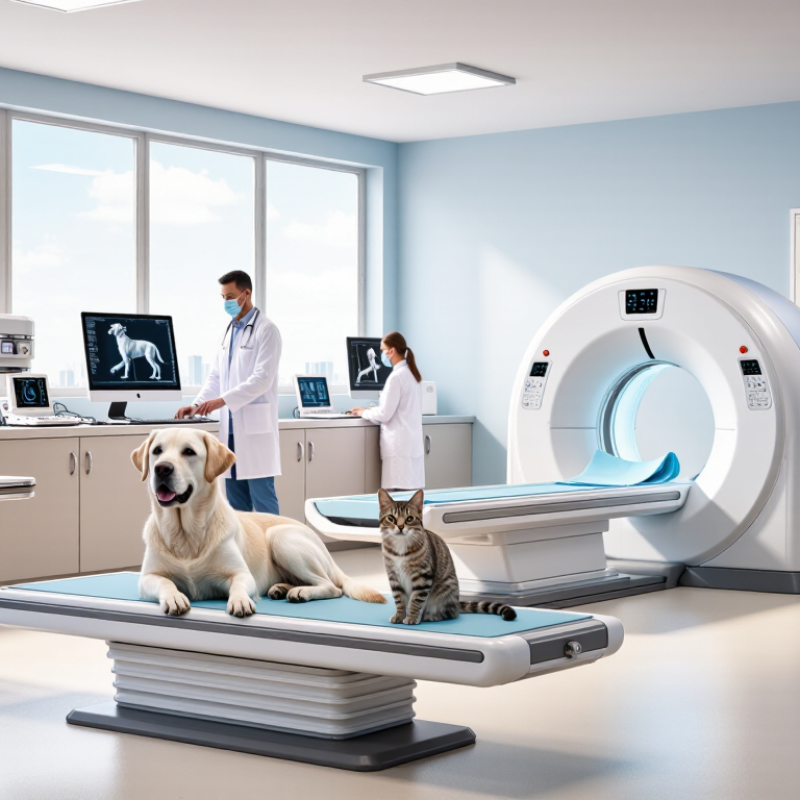
Advanced imaging should begin with a clear clinical question, not a machine. Ask what you need to confirm, exclude, or locate. “Why is my dog limping?” is too broad. “Is there a tendon tear in the right shoulder?” guides a more useful decision. The American College of Veterinary Radiology supports an evidence-based process that connects history, examination, and imaging findings.
Start with the patient’s age, symptoms, physical findings, and likely diseases. A cat with repeated vomiting may need abdominal radiographs before more advanced testing. A neurological examination can help determine whether imaging should focus on the brain, spine, or peripheral nerves. Choose the modality that best answers the question. Consider image quality, radiation exposure, contrast requirements, sedation, cost, and local expertise.
A good imaging plan also asks how results will change treatment. If surgery is unlikely, an invasive test may add little value. If infection, bleeding, or cancer is possible, timing becomes important. Experienced clinicians should still challenge their first assumption, because familiar symptoms can hide uncommon conditions. I have seen imaging requests become clearer after one extra question: “What decision will this result support?” Radiologists and veterinarians can then review the images together, compare them with the examination, and explain limitations honestly. No scan replaces clinical judgment.
Advanced diagnostic imaging should begin with the clinical question, not the machine. A painful limb may need radiographs, while suspected internal bleeding could require CT. Both use ionizing radiation, but they are not interchangeable.
The FDA generally places diagnostic X-ray systems and computed tomography systems in Class II, yet they follow different equipment standards and product classifications. This distinction does not mean one modality is automatically safer. It reflects different designs, controls, and intended uses. The FDA estimates a typical adult chest X-ray at about 0.1 mSv, while an abdominal CT may reach approximately 8 mSv. These figures are human references, not fixed veterinary doses. A small dog, a large cat, and a sedated rabbit will not receive identical exposure.
That difference matters. CT collects many X-ray measurements around the patient, producing detailed cross-sectional images. It can reveal tiny lung nodules, complex fractures, or hidden abdominal lesions. A standard radiograph is faster, cheaper, and often sufficient for bones or the chest. Veterinary teams should review body size, scan length, repeat imaging, and sedation needs. The American College of Radiology emphasizes dose optimization through appropriate protocols, not simply choosing the lowest setting. The IAEA also recommends using diagnostic reference levels to identify unusually high exposure. Those benchmarks are useful, but veterinary reference data remain less complete. A single number can mislead. Ask what information the scan will change.
How to Choose Advanced Diagnostic Imaging for Pets?
Match anatomy and tissue type to the test, not the equipment. Ultrasound works well for fluid-filled organs and moving soft tissues. It can show gallbladder sludge, intestinal layering, or a bladder stone during a live examination. It avoids ionizing radiation, but gas, bone, and body size can limit the view. A 2022 American Veterinary Medical Association report estimated 65.1 million U.S. households owned dogs and 46.5 million owned cats. With this clinical volume, careful test selection matters.
CT is usually stronger for bone, lungs, nasal passages, and complex trauma. It produces fast, cross-sectional images and often requires sedation or anesthesia. MRI offers greater soft-tissue contrast for the brain, spinal cord, joints, and nerves. It takes longer and may reveal findings that do not explain the animal’s pain. That is where judgment matters. More detail is not always better care.
A practical case shows the difference. A cat with repeated vomiting may need ultrasound to assess intestinal layers and motility. A dog with suspected nasal cancer may benefit from CT for bone invasion. A puppy with hind-limb weakness may need MRI of the spine. Professional interpretation remains essential, because image quality, positioning, and patient history can change the answer. Mistakes happen when clinicians choose a modality before identifying the tissue involved.
How to Choose Advanced Diagnostic Imaging for Pets?
Assess Anesthesia Risk Under AAHA 2020 Monitoring Guidelines
Advanced imaging can reveal hidden disease, but anesthesia risk deserves equal attention. A quiet cat may still have heart, kidney, or airway problems. The veterinary team should review history, age, medications, hydration, and previous anesthetic reactions. A focused physical examination follows. Blood tests, blood pressure, or cardiac testing may be appropriate for selected patients.
The 2020 anesthesia monitoring guidance emphasizes preparation, trained supervision, and continuous assessment. Before imaging, confirm working oxygen, ventilation, suction, emergency drugs, and warming equipment. During anesthesia, monitor oxygenation, ventilation, circulation, temperature, and anesthetic depth. Use appropriate sensors and record measurements at regular intervals. One trained person should remain responsible for the patient, not the scan alone.
Details matter.
For a short scan, risk may seem small. It is not automatically small. Positioning can restrict breathing, especially in obese or brachycephalic animals. An MRI environment may also limit physical access during the procedure. Discuss fasting, airway control, pain, recovery location, and discharge criteria before consent. Recovery needs the same vigilance as induction. Many complications appear after the images are finished.
No checklist catches everything. In practice, teams sometimes focus too heavily on image quality and underestimate recovery time. That deserves honest review. Ask whether the scan will change treatment, whether another method could answer the question, and whether the facility can support this patient’s specific risks. The safest choice is the one matched to the animal, the equipment, and the team’s monitoring capability.
| Assessment Dimension | Lower-Risk Indicators | Moderate- to High-Risk Indicators | Imaging and Anesthesia Planning Implications | AAHA 2020 Monitoring Considerations |
|---|---|---|---|---|
| Clinical urgency | Stable patient; imaging can be scheduled after optimization. | Trauma, respiratory distress, suspected intracranial disease, shock, or rapidly worsening signs. | Choose the fastest diagnostic pathway that answers the clinical question. Stabilize life-threatening abnormalities before non-emergency imaging whenever possible. | Assign a dedicated trained individual to monitor the patient continuously and record findings at appropriate intervals. |
| Need for anesthesia or deep sedation | Some radiographs or ultrasound examinations may be completed with gentle restraint or minimal sedation. | Motion-sensitive MRI, many CT studies requiring immobility, painful positioning, or procedures requiring airway control. | Use the least invasive technique that can produce diagnostic-quality images. Do not select an imaging modality solely to avoid anesthesia if it cannot answer the medical question. | Monitoring should be matched to anesthetic depth and patient risk; pulse oximetry alone does not assess ventilation adequately. |
| Airway and respiratory status | Normal respiratory effort, clear or stable lung findings, and no evidence of upper-airway obstruction. | Dyspnea, brachycephalic anatomy, laryngeal disease, pneumonia, pulmonary edema, or increased aspiration risk. | Consider whether the patient can safely tolerate positioning, transport, sedation, and recovery. Plan airway access and oxygen support before leaving the treatment area. | Monitor oxygenation with pulse oximetry and ventilation with end-tidal carbon dioxide when available and clinically appropriate. Observe respiratory rate, pattern, and effort. |
| Cardiovascular status | Stable perfusion, normal or controlled heart rate, acceptable blood pressure, and no clinically important arrhythmia. | Hypotension, poor perfusion, significant arrhythmia, decompensated heart disease, severe anemia, or suspected shock. | Correct major perfusion abnormalities before elective imaging. Consider shorter procedures, enhanced staffing, and a setting equipped for cardiovascular support. | Monitor heart rate and rhythm, pulse quality, mucous membranes, capillary refill time, and blood pressure. ECG assists rhythm assessment but does not measure perfusion. |
| Renal and hepatic function | No clinical evidence of major organ dysfunction and acceptable recent laboratory results when indicated. | Azotemia, oliguria, dehydration, hepatic dysfunction, electrolyte abnormalities, or suspected toxin exposure. | Review whether contrast is necessary. Correct dehydration and important electrolyte disturbances when feasible, and individualize drug selection and dosing. | Document the anesthetic plan, response to treatment, fluid therapy, and recovery observations. Reassess abnormal physiology rather than relying only on a single test result. |
| Patient age and body condition | Healthy adult with stable body condition. | Neonatal or geriatric patient, very small patient, obesity, cachexia, or impaired thermoregulation. | Adjust drug doses and warming strategies to the patient. Minimize fasting-related risk and avoid prolonged positioning or recovery delays. | Monitor body temperature during anesthesia and recovery; provide active warming or cooling as indicated and prevent thermal injury. |
| Neurologic status | Normal mentation and stable neurologic examination. | Altered mentation, seizures, increased intracranial pressure concern, severe head trauma, or impaired protective reflexes. | Prioritize stabilization, oxygenation, ventilation, and careful positioning. MRI may provide superior soft-tissue detail, while CT is often faster for bone, acute trauma, and some emergency evaluations. | Track anesthetic depth, ventilation, oxygenation, blood pressure, temperature, and recovery quality; prevent hypoventilation and delayed recognition of deterioration. |
| Contrast-agent considerations | No known previous contrast reaction and no major contraindicating condition. | Previous contrast reaction, severe renal compromise, dehydration, unstable cardiovascular status, or inability to protect the airway. | Confirm that contrast will change case management. Review history, indication, hydration status, and emergency preparedness before administration. | Continue observation after contrast administration and be prepared to manage acute adverse reactions, including respiratory or cardiovascular compromise. |
| Patient movement and positioning | Comfortable, cooperative patient that can maintain the required position. | Severe pain, fractures, spinal instability, respiratory compromise in dorsal recumbency, or inability to remain still. | Provide analgesia and use positioning aids. Avoid unnecessary manipulation and select the modality that provides adequate information with the least destabilizing positioning. | Recheck circulation, ventilation, pressure points, and body temperature after repositioning. Maintain continuous observation during transport and scanning. |
| Monitoring and staffing readiness | Appropriately trained personnel, functioning equipment, documented plan, and a quiet recovery area are available. | No dedicated monitor, inadequate emergency equipment, unreliable monitoring devices, or recovery space not prepared. | Delay elective imaging until essential resources are ready. For high-risk patients, use a facility capable of advanced airway, oxygen, cardiovascular, and emergency support. | AAHA guidance emphasizes continuous patient observation, appropriate physiologic monitoring, accurate documentation, equipment checks, and active monitoring through recovery. |
| Post-anesthetic recovery | Patient is warm, breathing adequately, responsive, and hemodynamically stable before discharge from monitored care. | Persistent hypothermia, hypoventilation, pain, dysphoria, hypotension, delayed awakening, or repeated vomiting. | Extend supervised recovery and provide oxygen, analgesia, airway support, or additional treatment as clinically indicated. Do not transfer responsibility before the patient is stable. | Continue monitoring until the patient is alert enough to maintain an airway and has stable respiratory and cardiovascular function. Record recovery events and interventions. |
| Overall risk classification | Lower risk Stable patient, limited comorbidity, predictable procedure, and complete monitoring resources. |
Moderate risk Controlled systemic disease, difficult positioning, contrast requirement, or prolonged imaging time. |
Higher risk Unstable physiology, serious respiratory or cardiovascular disease, major neurologic compromise, or urgent imaging during incomplete stabilization. |
Risk classification should guide preparation, staffing, monitoring intensity, recovery location, and escalation decisions. It should be reassessed whenever the patient's condition changes. |
This table is a clinical planning aid, not a substitute for an individualized examination, informed consent, local protocols, or professional veterinary judgment. The attending veterinarian should determine the safest imaging modality, sedation or anesthesia plan, monitoring requirements, and recovery pathway for each patient.
Clear images do not guarantee a clear diagnosis. Ask whether an ACVR board-certified veterinary radiologist will interpret the study. ACVR diplomates complete specialized training and pass examinations in diagnostic imaging. Their report should identify image limitations, not hide them.
Look for thin-slice CT data, appropriate MRI sequences, and complete patient positioning. A 2022 review in Veterinary Radiology & Ultrasound linked interpretation quality to technical image quality, clinical history, and reader expertise. These factors work together. One missing detail can change the reading.
Ask for the radiologist’s credentials and a written report. The report should explain findings, likely causes, and recommended next steps. It should also state when results are uncertain. That honesty matters.
The American College of Radiology reports that human radiology interpretation discrepancies commonly range from 3% to 5%; veterinary rates may differ because published data remain limited. This comparison is imperfect, but it shows why independent review matters. A second look can catch subtle lung nodules, small spinal lesions, or early joint changes. I would not choose equipment by marketing language alone. I would ask who reads the images, how quickly, and whether the clinical team can discuss confusing findings.
A clear question connects symptoms, examination findings, and the needed scan. “Is the right shoulder tendon torn?” is useful. “Why is my dog limping?” is too broad.
Age, symptoms, and physical findings help identify the body area and likely disease. Repeated vomiting may begin with abdominal radiographs. A neurological examination may direct imaging toward the brain, spine, or nerves.
Radiographs are fast, affordable, and often suitable for bones or the chest. They may answer a simple fracture question without advanced testing. Sometimes, simple is better.
CT creates cross-sectional images from many X-ray measurements. It can show small lung nodules, complicated fractures, or hidden abdominal lesions. It may help when ordinary radiographs cannot answer the question.
No. Both use ionizing radiation, but CT collects more measurements around the patient. A typical adult chest radiograph is about 0.1 mSv. An abdominal CT may be about 8 mSv. These are human references, not fixed pet doses.
Body size, scan length, repeated images, and the selected protocol all matter. A small dog, large cat, and rabbit will not receive identical exposure. Dose should be optimized, not guessed from one number.
Discuss image quality, sedation, contrast needs, cost, local expertise, and possible radiation exposure. Ask how the result will change treatment. If surgery is unlikely, an invasive test may add little value.
No scan tells the whole story. Veterinary teams should compare images with examination findings. They should explain limitations honestly. I might question my first assumption. Familiar symptoms can hide uncommon conditions.
Advanced diagnostic imaging for pets begins with a clearly defined clinical question, allowing veterinarians to select the most appropriate examination rather than relying on technology alone. By applying an evidence-based approach, the care team can compare the benefits and limitations of ultrasound, CT, and MRI according to the suspected condition, body region, and tissue type. Understanding the difference between imaging methods that use ionizing radiation is also essential when discussing safety, diagnostic value, and appropriate case selection.
The question “what are the benefits of advanced diagnostic imaging for pets” can be answered through more accurate disease detection, improved treatment planning, and better-informed medical decisions. Before imaging, veterinarians should assess anesthesia needs and patient risk while following structured monitoring principles throughout the procedure. Finally, image quality and interpretation should be verified by an appropriately qualified veterinary imaging specialist, helping ensure that findings are reliable, clinically relevant, and clearly communicated to support the pet’s care.

WellData Medical